Late-in-life Exercise Training Increases Intracellular Protein Recycling in the Heart

Graphic: Late-in-life Exercise Training Increases Intracellular Protein Recycling in the Heart
Exercise induces autophagy, a “garbage recycling system” that clears damaged cellular components, in the aging heart

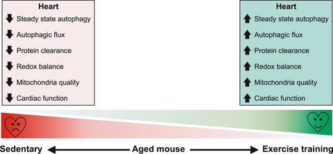
Heart cells (known as myocytes) work hard. Over a human lifetime, the heart beats approximately 2.5 billion times. As myocytes age, and especially in the presence of disease, they accumulate damaged intracellular components such as misfolded proteins. This build-up of damaged cellular material can cause cardiac dysfunction, diminish quality of life, and lead to premature death.

Cells have developed a means of identifying and recycling damaged components, through a mechanism termed autophagy (auto=‘self’; phagy=‘eating’). However, these autophagic control processes decline with aging—precisely when heart cells need them the most. Nutrition and Integrative Physiology PhD student Jaemin Cho, together with University of Utah Health researchers J. David Symons, PhD, and Sihem Boudina, PhD, collaborated on an investigation into whether late-in-life exercise in mice enhanced the protein recycling process. They observed that three months of daily treadmill training in very old mice dramatically increased autophagic machinery, enhanced the clearance of damaged proteins, and improved cardiac function. These findings provide the first evidence that late-in-life exercise training can improve cardiomyocyte quality control and function.

References:

Issues Icon

Late-in-life treadmill training rejuvenates autophagy, protein aggregate clearance, and function in mouse hearts. Cho JM, Park SK, Ghosh R, Ly K, Ramous C, Thompson L, Hansen M, Mattera MSLC, Pires KM, Ferhat M, Mookherjee S, Whitehead KJ, Carter K, Buffolo M, Boudina S, Symons JD. Aging Cell. 2021 Oct;20(10):e13467.

A Cellular Structure that Protects Against Amino Acid Stress

A Cellular Structure that Protects Against Amino Acid Stress
MDCs are membrane-bound structures that form from mitochondria in response to elevated amino acid levels in cells. MDCs cooperate with vacuoles (organelles containing digestive enzymes) to protect cells from amino acid toxicity.

Amino acids form the basic building blocks of all life, used by cells both as fuel and in building proteins and other complex molecules. Cellular amino acid levels must be tightly controlled. Amino acid surplus is problematic and is a characteristic of many age-related diseases, including cancer and diabetes. Scientists do not yet fully understand how cells detect and respond to amino acid excess, and how this excess causes cellular damage. Research from University of Utah Health scientist Adam Hughes, PhD, and colleagues discovered a new mechanism by which cells protect themselves from the toxic effects of amino acid excess. When faced with high amino acid levels, cells generate a membrane-bound structure from mitochondria, called the mitochondrial-derived compartment (MDC). MDCs promote the breakdown of amino acids. Loss of MDCs, in combination with other systems that regulate amino acid levels, causes cells to become highly sensitive to deviations in amino acid supply, leading to cell death. These results shed light on how cells regulate nutrient metabolism, advancing our understanding of how cells maintain metabolic stability.

References:

Issues Icon

Mitochondrial-derived compartments facilitate cellular adaptation to amino acid stress. Schuler MH, English, AM, Xiao T, Campbell TJ, Shaw JM, Hughes AL. Molecular Cell. 2021. Sep 16;81(18):3786-3802.e13.

Press Releases and Media:

U of U Health Key Faculty Collaborators:

Janet Shaw, PhD

MIC-Drop: A New Technology to Accelerate Gene Discovery

Graphic: MIC-Drop: A New Technology to Accelerate Gene Discovery
Hundreds of colored MIC-Drop droplets, each one-billionth of a liter in volume, are loaded into a microscopic glass needle in preparation for injection into zebrafish embryos. Each droplet contains the molecular machinery needed to edit a different zebrafish gene, along with a DNA “barcode” that can easily be read to identify which gene the droplet will edit.

Genome editing methods, such as the Nobel prize–winning CRISPR technology, enable scientists to make precise changes to genetic code. Beyond the potential to correct disease-causing mutations in humans, researchers discover genes’ functions by disrupting targeted genes in flies, fish, and other organisms and observing the effects on health and behavior. However, until recently, only one gene could be targeted at a time.

University of Utah Health researchers from the labs of Joseph Yost, PhD, and Randall Peterson, PhD, recently developed a method to target thousands of genes in parallel, rapidly accelerating discovery. Their Multiplexed Intermixed CRISPR Droplets (MIC-Drop) technique packages CRISPR components into tiny, oil-encased droplets, which each include a DNA “barcode” and can mingle without exchanging contents. By injecting a droplet into an organism, researchers can simultaneously edit a gene and mark the animal with easily recoverable information about which gene was targeted. The team has used MIC-Drop to identify sixteen genes that are critical for healthy heart development and function, and expects it to continue to accelerate the discovery of gene functions.

References:

Issues Icon

MIC-Drop: A platform for large-scale in vivo CRISPR screens. Parvez S, Herdman C, Beerens M, Chakraborti K, Harmer ZP, Yeh JJ, MacRae CA, Yost HJ, Peterson RT. Science. 2021 Sep 3;373(6559):1146-1151.

Press Releases and Media:

Focused Ultrasound as a Non-invasive Treatment for Breast Cancer

Graphic- Focused Ultrasound as a Non-invasive Treatment for Breast Cancer
Pictures of the non-invasive breast cancer treatment technology using a breast-specific magnetic resonance-guided, focused ultrasound system and real-time magnetic resonance image monitoring. A) Breast magnetic resonance-guided focused-ultrasound system installed in a magnetic resonance imaging scanner. B) Treatment cylinder component of the system where the treated breast is placed during the procedure. C) Temperature rise in the breast tumor during the ablation procedure. D) Cumulative thermal dose measured in cumulative equivalent minutes at 43°C (CEM 43°C) during the treatment, with red showing where cancer cells have been destroyed.


As physicians discover breast cancers at earlier stages, many women seek therapies that are effective yet non-invasive and non-scarring. Allison Payne, PhD, and her colleagues have developed a magnetic resonance-guided, focused ultrasound system specifically designed for breast tumor therapy. Steered by the physician, this system delivers high-intensity ultrasound waves to a precise area inside the breast, where non-invasively destroy malignant tissues are non-invasively destroyed with heat. This system was designed specifically for the patient’s comfort, with her laying on a customized table and her breast suspended in a treatment cylinder. Comfort is important because the patient may be inside the MRI machine for more than an hour. An additional innovation was the development of magnetic resonance imaging techniques that monitor, in real time, the temperature increase caused by the high-intensity ultrasound within the treatment tissues. This system is currently undergoing clinical trials and is with a goal of ultimately gaining FDA approval.  For patients who are medically-eligible, this technology represents a significant advance in breast cancer therapy.

References:

Issues Icon

Design and characterization of a laterally mounted phased-array transducer breast-specific MRgHIFU device with integrated 11-channel receiver array. Payne, A, Merrill R, Minalga E, Vyas U, de Bever J, Todd N, Hadley R, Dumont E, Neumayer L, Christensen D, Roemer R, Parker D. Med Phys. 2012 March;39(3):1552.

Issues Icon

An 11-channel radio frequency phased array coil for magnetic resonance guided high-intensity focused ultrasound of the breast. Minalga E, Payne A, Merrill R, Todd N, Vijayakumar S, Kholmovski E, Parker DL, Hadley JR. Magn Reson Med. 2013 January;69(1):295.

Issues Icon

In vivo evaluation of a breast-specific magnetic resonance guided focused ultrasound system in a goat udder model.Payne A, Todd N, Minalga E, Wang Y, Diakite M, Hadley R, Merrill R, Factor R, Neumayer L, Parker DL. Med Phys. 2013 July;40(7):073302.

Issues Icon

A breast-specific MR guided focused ultrasound platform and treatment protocol: first-in-human technical evaluation. A  Payne A, Merrill R, Minalga E, Hadley JR, Odéen H, Hofstetter LW, Johnson S, Tunon de Lara C, Auriol S, Recco S, Dumont E, Parker DL, Palussiere J. IEEE Trans Biomed Eng. 2021 Mar;68(3):893.

Press Releases and Media:

U of U Health Key Faculty Collaborators

Dennis L. Parker, PhD
J. Rock Hadley, PhD
Henrik Odéen, PhD

Glucagon Be-Gone: A Cure for Type 1 Diabetes?

Glucagon Be-Gone: A Cure for Type 1 Diabetes?
A re-grown pancreatic islet in a previously diabetic mouse treated with antagonists to glucagon action.

Cells within the pancreas produce the hormones insulin and glucagon, which have opposing actions on blood glucose. Insulin decreases blood glucose levels by promoting the utilization and storage of glucose while simultaneously repressing glucagon secretion. Glucagon opposes these actions, stimulating glucose production by the liver. In type 1 diabetes, where the insulin-producing cells are destroyed, glucagon levels remain perpetually elevated. The increase in glucagon is an important—but underappreciated—contributor to diabetes and its complications.

University of Utah Health investigator William Holland, PhD, and colleagues found that blocking glucagon action in mice restored levels of both insulin and glucose, effectively curing type 1 diabetes. Their groundbreaking work, which was published in the Proceedings of the National Academy of Sciences, reveals that suppressing glucagon production or action may be a viable means of treating type 1 diabetes.

References:

Issues Icon

Glucagon blockade restores functional β-cell mass in type 1 diabetic mice and enhances function of human islets. Wang MY, Dean ED, Quittner-Strom E, Zhu Y, Chowdhury KH, Zhang Z, Zhao S, Li N, Ye R, Lee Y, Zhang Y, Chen S, Yu X, Leonard DC, Poffenberger G, Von Deylen A, McCorkle SK, Schlegel A, Sloop KW, Efanov AM, Gimeno RE, Scherer PE, Powers AC, Unger RH, Holland WL. Proceedings of the National Academy of Sciences of the United States of America. 2021 Mar 2;118(9):e2022142118.

Press Releases and Media:

Controlling the Spread of Antibiotic Resistance

Graphic- Controlling the Spread of Antibiotic Resistance
Illustration of the effect of contact precautions (CPe) on the baseline infection transmission rate (Inf), comparing patients on contact precautions mandatory gowns, gloves, and masks; left) with those not on contact precautions (right).

Healthcare-associated infections due to antibiotic-resistant bacteria are costly and deadly. Michael Rubin, MD, and Matthew Samore, MD, generated new evidence on the effect of infection-prevention practices on the transmission of antibiotic-resistant pathogens, particularly methicillin-resistant Staphylococcus aureus (MRSA). They elucidated the epidemiological mechanisms for the progressive decline in rates of MRSA colonization and infection following the nationwide implementation of a program at Veterans Affairs healthcare facilities. The program introduced active surveillance to detect MRSA colonization in patients and placement of colonized patients on contact precautions, which involve the use of gowns and gloves by visiting healthcare workers. They used rigorous methods to evaluate the impact of this program and to model its cost-effectiveness, showing that the decrease in MRSA transmission that occurred following introduction of the program was largely due to contact precautions. Further, implementation of the program likely prevented a substantial number of infections from non-MRSA organisms, and was, therefore, highly cost-effective. Their studies generated important evidence for the role of contact precautions in preventing healthcare-associated infections, an issue of critical importance in healthcare epidemiology.

References:

Issues Icon

Relationships between the importation, transmission, and nosocomial infections of methicillin-resistant Staphylococcus aureus: an observational study of 112 Veterans Affairs Medical Centers. Jones M, Ying J, Huttner B, Evans M, Maw M, Nielson C, Rubin MA, Greene T, Samore MH. Clin Infect Dis. 2014 January;58(1):32.

Issues Icon

Efficient parameter estimation for models of healthcare-associated pathogen transmission in discrete and continuous time. Thomas A, Redd A, Khader K, Leecaster M, Greene T, Samore M. Math Med Biol. 2015 March;32(1):79.

Issues Icon

Economic analysis of Veterans Affairs initiative to prevent methicillin-resistant Staphylococcus aureus infections. Nelson RE, Stevens VW, Khader K, Jones M, Samore MH, Evans ME, Douglas Scott, R 2nd, Slayton RB, Schweizer ML, Perencevich EL, Rubin MA. Am J Prev Med. 2016 May;50(5 Suppl 1):S58.

Issues Icon

Vital Signs: Trends in Staphylococcus aureus Infections in Veterans Affairs Medical Centers – United States, 2005-2017. Jones M, Jernigan JA, Evans ME, Roselle GA, Hatfield, KM, Samore MH. MMWR Morb Mortal Wkly Rep. 2019 March 8;68(9):220.

Issues Icon

Variation and trends in transmission dynamics of methicillin-resistant Staphylococcus aureus in Veterans Affairs hospitals and nursing homes. Khader K, Thomas A, Jones M, Toth D, Stevens V, Samore MH, the CDC Modeling Infectious Diseases in Healthcare Program. Epidemics. 2019 September;28:100347.

Issues Icon

Expanding an economic evaluation of the Veterans Affairs (VA) methicillin-resistant Staphylococcus aureus (MRSA) prevention initiative to include prevention of infections from other pathogens. Nelson RE, Goto M, Samore MH, Jones M, Stevens VW, Evans ME, Schweizer ML, Perencevich EN, Rubin MA. Clin Infect Dis. 2021 January 29;72(Supplement_1):S50.

Issues Icon

National estimates of healthcare costs associated with multidrug-resistant bacterial infections among hospitalized patients in the United States. Nelson RE, Hatfield KM, Wolford H, Samore MH, Scott RD, Reddy SC, Olubajo B, Paul P, Jernigan JA, Baggs J. Clin Infect Dis. 2021 January 29;72(Supplement_1):S17.

Issues Icon

Association between contact precautions and transmission of methicillin-resistant Staphylococcus aureus in  Veterans Affairs hospitals. Khader K, Thomas A, Stevens V, Visnovsky L, Nevers M, Toth D, Keegan LT, Jones M, Rubin M Samore MH. JAMA Netw Open. 2021 March 1;4(3):e210971.

A New Form of Glutamate Signaling Discovered in Migraine

Graphic- A New Form of Glutamate Signaling Discovered in Migraine
A) Plumes result from an imbalance between release of glutamate from neurons and its reuptake by astrocytes. B) Plumes occur both during initiation (left) and propagation (right) of spreading depolarizations (SD), waves of excitation that occur in multiple neurologic diseases.

Migraine is a disorder of the sensory nervous system, consisting of head pain and debilitating sensory amplifications, wherein the normal senses we use in daily living become painful.  Although migraine is thought to be the result of increased nervous system excitability, the actual mechanisms are poorly understood.  Using two-photon microscopy, K.C. Brennan, MD, and colleagues discovered unusual activity of the excitatory neurotransmitter glutamate in mice carrying a mutated gene identified from a family with inherited migraine. These ‘plumes’ of glutamate release were much more common in mice carrying the migraine mutation than in those without.  Moreover, a flurry of plume events preceded spreading depolarizations, massive waves of excitation that cause the migraine aura.  This novel finding is important beyond migraine, because spreading depolarizations occur in several other conditions, including stroke and traumatic brain injury. Thus, plumes represent a novel mechanism of excitability in the brain, relevant to a broad array of neurologic diseases.

References:

Issues Icon

Non-canonical glutamate signaling in a genetic model of migraine with aura. Parker PD, Suryavanshi PS, Melone M, Sawant-Pokam PM, Reinhart KM, Kaufmann D, Theriot JJ, Pugliese A, Conti F, Shuttleworth CW, Pietrobon D, Brennan KC. Neuron.2021 Feb 17;109(4):611.e8. PMID: 33321071. 

Press Releases and Media:

U of U Key Faculty Collaborators

Punam Sawant-Pokam, PhD
Dan Kaufmann, PhD

The Levonorgestrel Intrauterine Device is Effective as Emergency Contraception

Graphic- The Levonorgestrel Intrauterine Device is Effective as Emergency Contraception

Emergency contraception prevents pregnancy after sexual intercourse. The FDA has approved two “morning-after” pills for this purpose, and the copper intrauterine device (IUD) is an order of magnitude more effective than these oral options. For non-emergency contraception, many people prefer the hormone levonorgestrel-releasing IUD over the copper IUD because it reduces or eliminates menstrual bleeding and cramping. 

University of Utah Health researcher David Turok, MD, and colleagues were the first to evaluate the levonorgestrel IUD for emergency use. Their NIH-funded research randomized 638 people seeking emergency contraception in six Utah clinics to either the levonorgestrel IUD or the copper IUD and found the hormonal IUD to be as effective as the copper IUD for emergency pregnancy prevention. As a result, levonorgestrel IUD has already been approved for use by all Planned Parenthood Federation of America clinics nationwide. 

By demonstrating that people can begin a levonorgestrel IUD at any time in the menstrual cycle, regardless of recent unprotected intercourse, this study introduced the first new emergency contraception option in a decade, providing effective contraception with a favorable side-effect profile.

References:

Issues Icon

Levonorgestrel vs. copper intrauterine devices for emergency contraception. Turok DK, Gero A, Simmons RG, Kaiser JE, Stoddard, GJ, Sexsmith CD, Gawron LM, Sanders JN. N Engl J Med. 2021 Jan 28;384(4):335-344.

Press Releases and Media:

U of U Health Key Faculty Collaborators

Rebecca Simmons, PhD
Jennifer Kaiser, MD
Lori Gawron, MD
Jessica Sanders, PhD

Effectiveness of Physical Therapy for Back Pain

Graphic- Effectiveness of Physical Therapy for Back Pain
A randomized controlled trial examined the effects of early referral to physical therapy plus a single educational session versus a single educational session alone on disability among patients with acute sciatica. Patients referred to physical therapy reported reduced pain and disability. Reproduced with permission from https://www.acpjournals.org/doi/10.7326/M20-4187. ©American College of Physicians.


Back pain is among the most common reasons for visits to physicians and physical therapists and is the costliest health condition in the US. Twenty years ago, the scientific basis to guide decision-making on treatments for specific types of back pain was weak. Since then, Julie Fritz, PhD, and colleagues have conducted a series of rigorous randomized clinical trials examining patients with acute back pain, back pain accompanied by sciatica, and back pain due to spinal stenosis. Their recent study found that patients with back pain and sciatica who were referred to physical therapy for treatment with exercise and manual therapy were more likely to rate their treatment as successful and reported significantly greater reductions in pain and disability than patients who were not referred. These studies have defined effective physical therapy treatments for these conditions and facilitated clinical decision making and strategies to personalize physical therapy treatments. 

References:

Issues Icon

Early physical therapy vs usual care in patients with recent-onset low back pain. Fritz JM, Magel JS, McFadden M, Asche CV, Thackeray A, Meier W, Brennan G. JAMA. 2015 October 14;314(14):1459.

Issues Icon

Prevention and treatment of low back pain: evidence, challenges, and promising directions. Foster NE, Anema JR, Cherkin D, Chou R, Cohen SP, Gross DP, Ferreira PH, Fritz JM, Koes BW, Peul W, Turner JA, Maher CG. Lancet. 2018 June 9;391(10137):2368.

Issues Icon

Physical therapy referral from primary care for acute back pain with sciatica: a randomized controlled trial. Fritz JM, Lane E, McFadden M, Brennan G, Magel JS, Thackeray A, Minick K, Meier W, Greene T. Ann Intern Med. 2021 January;174(1):8.

Press Releases and Media:

U of U Health Key Faculty Collaborators

Jake Magel, PhD
Tom Greene, PhD
Anne Thackeray, PhD
Elizabeth Lane, PhD

Myocardial Recovery in Chronic Heart Failure

Graphic- Myocardial Recovery in Chronic Heart Failure
The recovering heart increases the production of a mitochondrial pyruvate carrier protein (MPC), which transports pyruvate into the mitochondria where it is oxidized and fuels energy production.

Chronic heart failure is a disease with poor prognosis and currently is a global epidemic. University of Utah Health investigator Stavros George Drakos, MD, and colleagues analyzed human heart tissue and produced evidence refuting the widely held notion that prolonged off-loading of the failing heart induced by cardiac assist devices results in disuse atrophy that further deteriorates heart function. In contrast, there was significant heart recovery following the implantation of cardiac assist devices. These beneficial outcomes were recently validated in a multicenter trial co-led by the University of Utah.

To elucidate the mechanisms driving these favorable outcomes, they performed microstructural and metabolic studies. They discovered that, in humans and animals, the recovering heart exhibited preference for glucose oxidation over lactate production. To accomplish this, the recovering heart increases the production of a mitochondrial pyruvate carrier protein (MPC), which transports pyruvate into the mitochondria where it is oxidized and fuels energy production. This research has been conducted through the multidisciplinary Utah Cardiac Recovery Program, comprising investigators from the Nora Eccles Harrison Cardiovascular Research & Training Institute, multiple U of U departments, Intermountain Healthcare, Salt Lake VA as well as international collaborators. These recent discoveries provide critical scientific and clinical insights and novel therapeutic targets for heart failure.

References:

Issues Icon

Magnitude and time course of changes induced by continuous-flow left ventricular assist device unloading in chronic heart failure: insights into cardiac recovery. Drakos SG, Wever-Pinzon O, Selzman CH, Stehlik J, Gilbert EM, Alharethi EM, Reid BB, Budge D, Movsesian M, Li DY, Kfoury AG (UCAR Investigators). J Am Coll Cardiol. 2013 May 14;61(19):1985.

Issues Icon

Myocardial atrophy and chronic mechanical unloading of the failing human heart: implications for ventricular assist devices-induced cardiac recovery. Diakos NA, Selzman CH, Sachse FB, Stehlik J, Kfoury AG, Reid BB, Miller DV, Salama ME, Fang JC, Drakos SG. J Am Coll Cardiol. 2014 October 14;64(15):1602.

Issues Icon

Evidence of glycolysis upregulation and pyruvate mitochondrial oxidation mismatch during mechanical unloading of the failing human heart: implications for cardiac reloading and conditioning. Diakos NA, Navankasattusas S, Abel ED, Rutter J, McCreath L, Ferrin P, McKellar SH, Richardson R, Deberardinis R, Kfoury AG, Selzman CH, Stehlik J, Fang JC, Li DY, Drakos SG. JACC Basic Transl Sci. 2016 October;1(6):432.

Issues Icon

Impact of heart failure etiology on the incidence of cardiac recovery during mechanical unloading: a prospective study from the utah cardiac recovery program. Wever-Pinzon J, Selzman CH, Wever-Pinzon O, Catino A, Kfoury AG, Stehlik J, Al- Sarie M, Stoddard G, McKellar S, Bonios MJ, Koliopoulou A, Fang JC, Drakos SG. J Am Coll Cardiol. 2016 October 18;68(16):1741.

Issues Icon

Sheet-like remodeling of the transverse tubular system in human heart failure impairs excitation-contraction coupling and functional recovery by mechanical unloading. Seidel T, Navankasattusas S, Ahmad A, Diakos NA, Xu WD, Tristani-Firouzi M, Bonios MJ, Taleb I, Li DY, Selzman CH, Drakos SG*, Sachse FB* (*Co-Corresponding authors). Circulation. 2017 April 25;135(17):1632.

Issues Icon

 Cardiac rotational mechanics as a predictor of myocardial recovery in heart failure patients undergoing chronic mechanical circulatory support. Bonios MJ, Koliopoulou A, Wever-Pinzon O, Taleb I, Xu W, Stehlik J, Kfoury AG, Horne BD, Selzman CH, Fang JC, Bax JJ, Drakos SG. Circulation: Cardiovascular Imaging. 2018 April;11(4):e007117.

Issues Icon

The human heart contains distinct macrophage subsets with divergent origins and functions. Bajpai G, Schneider C, Bredemeyer A, Hulsman M, Nahrendorf MP, Epelman S, Kreisel D, Itoh A, Selzman C, Sankar T, Drakos SG, Lavine KJ. Nature Medicine. 2018 August; 24(8):1234.

Issues Icon

The role of non-glycolytic glucose metabolism in myocardial recovery upon mechanical unloading and circulatory support in chronic heart failure. Badolia R, Ramadurai D, Abel ED, Ferrin P, Taleb I, Sankar T, McKellar S, Yin M, Kfoury AG, Wever-Pinzon O, Fang JC, Selzman CH, Chaudhuri D, Rutter J, Drakos SG. Circulation. 2020 July 21;142(3):259.

Issues Icon

A prospective multicentre study of myocardial recovery using left ventricular assist devices (remission from Stage D heart failure: RESTAGE-HF): medium term and primary endpoint results. Birks EJ*, Drakos SG* (*co-corresponding authors), Patel SR, Lowes BD, Selzman CH, Starling RC, Trivedi J, Slaughter MS, Goldstein D, et al. Circulation. 2020 November 24;142(21):2016.

Issues Icon

The pyruvate-lactate axis modulates cardiac hypertrophy and heart failure. Cluntun AA, Badolia R, Lettlova S, Parnell KM, Shankar TS, Diakos NA, Olson KA, Taleb I, Tatum SM, Berg JA, Cunningham CC, Krokidi AT, Skedros S, Navankasattusas S, Cox JE, Ducker GS, Holland WL, McKellar SH, Rutter J, Drakos SG. Cell Metabolism. 2021 Mar 2;33(3):629.

Press Releases and Media

U of U Health Key Faculty Collaborators:

Craig H. Selzman, MD
Jared P. Rutter, PhD
Omar WeverPinzon, MD
Frank Bernd Sachse, Dr.-Ing

Reconstituting HIV Replication in a Test Tube

Graphic: Reconstituting HIV Replication in a Test Tube
Summary of the stepwise reconstitution of HIV-1 capsid-dependent replication and integration in a cell-free system. Credit: Janet Iwasa

Reverse transcription and integration are key events in retrovirus replication and are also targets of successful anti-HIV therapies. Reverse transcription creates a double-stranded DNA copy of the viral RNA genome, and integration archives that copy within the genome of the infected cell. However, studies of the mechanisms underlying these steps of the viral life cycle remain challenging because these processes are performed by viral core particles located deep within the infected cell cytoplasm and nucleus. To address this limitation, University of Utah Health investigator Wesley Sundquist, PhD, and colleagues reconstituted efficient HIV reverse transcription and integration in a cell-free system. They showed that the system responds appropriately to antiviral compounds. They also discovered that the viral capsid (the protein shell of a virus) plays an active role in supporting efficient reverse transcription. Thus, the entire core particle, including the outer capsid shell, is the true viral “replication complex”. This cell-free system is expected to enable new systematic analyses of viral replication and integration and thus help shed light on the first half of the viral life cycle.

References:

Issues Icon

Reconstitution and visualization of HIV-1 capsid-dependent replication and integration in vitro. Christensen DE, Ganser-Pornillos BK, Johnson JS, Pornillos O, Sundquist WI. Science. 2020 Oct;370(6513):eabc8420.

Press Releases and Media:

U of U Health Key Faculty Collaborators

Jarrod Johnson, PhD
Barbie Ganser-Pornillos, PhD
Owen Pornillos, PhD

Megakaryocytes and Platelets in Immune and Inflammatory Responses and in COVID-19

Viral Infections Alter Megakaryocyte and Platelet Gene Expression and Function. SARS-CoV-2 infection generations thromboinflammatory agonists which alter platelet gene expression and function to promote increased platelet-leukocyte aggregates and platelet reactivity.
SARS-CoV-2 infection generations thromboinflammatory agonists which alter platelet gene expression and function to promote increased platelet-leukocyte aggregates and platelet reactivity (Blood 2020).

Platelets—small cells which circulate in abundance in the bloodstream—are traditionally known for their ability to form clots and stop bleeding. Recent studies, however, have shown that platelets and their parent cells, megakaryocytes, also play a role in inflammation and infection. University of Utah Health investigators Robert Campbell, PhD, and Matthew Rondina, MD, and colleagues discovered that platelets and megakaryocytes respond robustly to infection, including COVID-19. These infection-driven changes in platelets activate clotting mechanisms and thus may contribute to the blood clots that complicate COVID-19 infection. 

Interestingly, Campell and Rondina and their collaborators also found that platelets and megakaryocytes demonstrate robust anti-viral defense properties that inhibit the spread of infection. Therefore, megakaryocytes from patients with genetic mutations in these anti-viral defense proteins are more susceptible to infection. These findings helped identify new pathways that regulate host responses to viral infection, and platelets are now being targeted therapeutically in patients with COVID-19.

References:

Issues Icon

Human megakaryocytes possess intrinsic antiviral immunity through regulated induction of IFITM3. Campbell RA, Schwertz H, Hottz ED, Rowley JW, Manne BK, Washington AV, Hunter-Mellado R, Tolley ND, Christensen M, Eustes AS, Montenont E, Bhatlekar S, Ventrone CH, Kirkpatrick BD, Pierce KK, Whitehead SS, Diehl SA, Bray PF, Zimmerman GA, Kosaka Y, Bozza PT, Bozza FA, Weyrich AS, Rondina MT. Blood. 2019 May 9;133(19):2013-2026.

Issues Icon

Platelet gene expression and function in COVID-19 patients. Manne BK, Denorme F, Middleton EA, Portier I, Rowley JW, Stubben CJ, Petrey AC, Tolley ND, Guo L, Cody MJ, Weyrich AS, Yost CC, Rondina MT, Campbell RA. Blood. 2020 Sep 10;136(11):1317-1329.

Issues Icon

COVID-19 patients exhibit reduced procoagulant platelet responses. Denorme F, Manne BK, Portier I, Petrey AC, Middleton EA, Kile BT, Rondina MT, Campbell RA. J Thromb Haemost. 2020 Nov;18(11):3067-3073.

Press Releases and Media:

U of U Health Key Faculty Collaborators

Hansjorg Schwertz, MD
Jesse W. Rowley, PhD
Paul F. Bray, MD
Guy A. Zimmerman, MD
Andrew S. Weyrich, PhD
Elizabeth A. Middleton, MD
Aaron C. Petrey, PhD
Christian Con Yost, MD

Visualizing the SARS-CoV-2 Life Cycle

Graphic: Visualizing the SARS-CoV-2 Life Cycle
3D model of SARS-CoV-2.

SARS-CoV-2 is defining disease of the current era. Many biological researchers have redirected their focus to understanding and defeating the virus, rapidly leading to new insights into how the virus gains access to and hijacks human cells. University of Utah Health investigator Janet Iwasa, PhD, and colleagues have used this information to create detailed molecular animations of different stages of the SARS-CoV-2 life cycle. The animations have been released to the research community and the public, embedded within a web-based user interface that allows users to interact with the animation in order to view annotations (such as protein names and citations), ask questions, and make comments. The annotation functionality, developed in collaboration with Miriah Meyer, PhD, now at Linköping University, is critical for describing the data used to create the visualization, and for discussing aspects of the life cycle that are not yet well understood.

References:

Press Releases and Media:

A Potent, Long-lasting HIV Capsid Inhibitor

Graphic: A Potent, Long-lasting HIV Capsid Inhibitor
The graph shows drug levels in people treated with single doses of Lenacapavir at different levels. The dotted red line shows the drug level required to inhibit HIV replication by 95%. The highest levels of Lenacapavir remain inhibitory for more than 24 weeks and are well tolerated.

Antiretroviral drugs have saved the lives of millions living with HIV/AIDS. However, problems with drug resistance limit available treatments, and inconsistent adherence to daily dosing schedules can lead to poor outcomes and new infections. Long-acting drugs that can overcome drug resistance by targeting new classes of viral proteins are therefore needed. University of Utah Health researcher Wesley Sundquist, PhD, and colleagues performed mechanistic studies and completed a phase 1 clinical trial of Lenacapavir, a molecule that inhibits the HIV capsid (outer shell). Lenacapavir is long-acting due to its high potency, slow release from the injection site, and slow clearance from the body. Single doses of Lenacapavir injected under the skin maintained antiviral concentrations for more than 6 months. The study validates therapies that target the HIV capsid, and demonstrates the potential of Lenacapavir as a long-acting agent to treat and prevent HIV infections. Lenacapavir was developed by Gilead Sciences, building on studies of HIV capsid structure and function from the Sundquist and Chris Hill, DPhil, laboratories (and others). Following a successful phase 3 trial, Lenacapavir has now been approved for use in Europe, and use in the US is pending FDA approval.

References:

Issues Icon

Clinical targeting of HIV capsid protein with a long-acting small molecule. Link JO, (Sundquist WI), et al. Nature. (2020) Aug;584(7822):614-618.

Press Releases and Media:

Supporting Health by Screening for Social Needs

Photo for "Supporting Health by Screening for Social Needs" entry, featuring a patient in an exam room being asked questions by a healthcare provider.

When patients struggle to get enough food, lack stable housing, or have limited access to transportation, their health can suffer. One analysis estimated that as much as 50 percent of the variation in health outcomes between counties in the U.S. can be attributed to social determinants of health like these. Such factors can limit the effectiveness of clinical care—but health care workers often don’t know what their patients are dealing with outside the clinic.

Addressing patients’ social needs is not always a priority in busy hospitals and clinics. But Andrea Wallace, PhD, RN, associate dean for research at the College of Nursing, has developed a way to make this a routine aspect of care. With systematic screening, patients’ unmet needs can be identified so clinicians can connect them with services that can help.

Wallace and her U or U Health colleagues have demonstrated that this screening can be done in emergency departments, which see disproportionate numbers of patients who are uninsured and have low incomes. The researchers are focused on finding the best ways to assess for social needs, considering factors such as language barriers, privacy concerns, and potential discomfort for patients and staff. In their most recent study, conducted at a children’s hospital in Salt Lake City, systematic screening allowed the team to identify 1,680 families with unmet social needs in a four-month period.

Wallace and colleagues have also shown how emergency departments can partner with a community service referral network to connect patients and families with services once needs have been identified. By helping secure access to healthy food, finding transportation to medical appointments, or assisting with rent and utilities, they can help patients get on a path to better health.

References:

Issues Icon

Social Needs Screening During Pediatric Emergency Department Visits: Disparities in Unmet Social Needs. Tedford NJ, Keating EM, Ou Z, Holsti M, Wallace AS, Robison JA. Acad Pediatr. 2022 Nov-Dec;22(8):1318-1327. doi: 10.1016/j.acap.2022.05.002.

Integrating social determinants of health screening and referral during routine emergency department care: evaluation of reach and implementation challenges. Wallace AS, Luther BL, Sisler SM, Wong B, Guo JW. Implement Sci Commun. 2021 Oct 7;2(1):114. doi: 10.1186/s43058-021-00212-y.

Implementing a Social Determinants Screening and Referral Infrastructure During Routine Emergency Department Visits, Utah, 2017-2018. Wallace AS, Luther B, Guo JW, Wang CY, Sisler S, Wong B. Prev Chronic Dis. 2020 Jun 18;17:E45. doi: 10.5888/pcd17.190339.

Neuronal Connections in the Retina

Graphic- Neuronal Connections in the Retina
A connectome from a normal rabbit retina (retinal connectome-1 or RC1 (top right) and a pathoconnectome from a diseased rabbit retina (Retinal Pathoconnectome-1 or RPC1 (top left). The image at bottom represents a “wiring diagram” showing the connections of 5113 neurons in a normal rabbit retina, connected by 8631 chemical and electrical synapses.

Understanding how neural systems are connected is a neuroscience grand challenge. The neural circuits in retina detect and process light information, then relay that information to the brain for further processing; the retina, therefore, provides a unique opportunity to understand how the brain processes information. Bryan Jones, PhD, and colleagues used electron microscopes to visualize the chemical and electrical synaptic connections that makes up the neural network. They further observed, in a transgenic rabbit model of early retinal degeneration, abnormal connectivity in the rod-photoreceptor network and novel synaptic connections derived from sprouting. 

These findings advanced understanding of retinal neuronal connections, the role of aberrant neuronal retinal wiring in blindness, and the potential development of new therapies to slow or reverse blindness. Since retinal wiring changes are analogous to other wiring changes within the brain, the retina also provides a more readily-accessible model to study other neural degenerative diseases, such as Alzheimer’s and Parkinson’s diseases. This work may also inform the development of new computational algorithms to improve artificial intelligence.

Bryan Jones, PhD, and Robert Marc, PhD
Bryan Jones, PhD, and Robert Marc, PhD.

References:

Issues Icon

Network architecture of gap junctional coupling among parallel processing channels in the mammalian retina. Sigulinsky CL, Anderson JR, Kerzner E, Rapp CN, Pfeiffer RL, Rodman TM, Emrich DP, Rapp KD, Nelson NT, Lauritzen JS, Meyer M, Marc RE, Jones BW. J. Neurosci. 2020 June 3;40(23):4483. PMID: 32332119 PMCID: PMC7275861

Issues Icon

A pathoconnectome of early neurodegeneration: Network changes in retinal degeneration. Pfeiffer RL, Anderson JR, Dahal J, Garcia JC, Yang J-H, Sigulinsky CL, Rapp K, Emrich DP, Watt CB, Johnstun HAB, Houser AR, Marc RE, Jones BW. Exp Eye Res. 2020 October;199:108196. PMID: 32810483 PMCID: PMC7554222

U of U Health Key Faculty Collaborator

Robert E. Marc, PhD

Unexpected Antiviral Activity of Spironolactone

Graphic- Unexpected Antiviral Activity of Spironolactone
A model of how the Epstein–Barr virus (EBV) uses xeroderma pigmentosum group B-complementing protein(XPB) to facilitate viral RNA synthesis by (1) initiating transcription from a viral gene promoter at the transcriptional start site (TSS) via RNA polymerase II (RNAPII) (viral pre-initiation complex (vPIC) required for viral transcription also present), after which; (2) SM binds to newly transcribed RNA, and then; (3) recruits XPB to facilitate elongation of the RNA transcript.

Epstein–Barr virus (EBV) is a human herpesvirus associated with clinical infections and several types of malignancies. Sankar Swaminathan, MD, and colleagues showed that a hypertension/heart failure drug, spironolactone, also has anti-EBV effects. The antiviral activity of spironolactone is not related to its mineralocorticoid blocking activity relevant to hypertension/heart disease, but, instead, to its ability to inhibit the function of a protein SM that is essential for EBV replication. In the first demonstration of its kind, this research also showed that EBV SM protein activates viral gene transcription by utilizing xeroderma pigmentosum group B-complementing protein (XPB) as a cofactor. 

This is the first demonstration that XPB is utilized as an essential cofactor by a human herpesvirus. The XPB protein is degraded by spironolactone. Therefore, XPB may be a useful therapeutic target to control EBV and other human herpesviruses. Further, because the SM protein acts at stages of the viral life cycle different from those targeted by available therapies, these observations pave the way for the development of novel anti-EBV drugs that address emerging drug resistance and toxicity problems. 

Sankar Swaminathan, MD, and Dinesh Verma, PhD.
Sankar Swaminathan, MD, and Dinesh Verma, PhD. Photo credit: Charlie Ehlert.

References:

Issues Icon

Spironolactone blocks Epstein–Barr virus production by inhibiting EBV SM protein function. Verma D, Thompson J, Swaminathan S. Proc Natl Acad Sci U S A. 2016 Mar 29;113(13):3609. Published online 2016 Mar 14. doi: 10.1073/pnas.1523686113

Issues Icon

Epstein–Barr virus co-opts TFIIH component XPB to specifically activate essential viral lytic promoters. Verma D, Church TM, Swaminathan S. Proc Natl Acad Sci U S A. 2020 June 9;117(23):13044. doi: 10.1 /pnas.2000625117

New Class of Therapy for Chronic Heart Failure

Graphic- New Class of Therapy for Chronic Heart Failure
Schematic illustration of microdomain organization of the structural protein BIN-1 within t-tubules of the adult ventricular cardiomyocytes. Originally published in Physiol Rev. 2017 January;97(1):227.


In the US, heart failure affects approximately eight million people, carries a poor prognosis, and represents the single largest healthcare cost for people over 65 years old. Most heart failure therapies rely on blocking the deleterious effects of stress hormones on the heart, but therapies aimed at repairing failing heart muscles are lacking. The lab directed by Nora Eccles Harrison Presidential Endowed Chair Robin Shaw, MD, PhD, and Nora Eccles Harrison Cardiovascular Research & Training Institute (CVRTI) investigators, have identified an architectural protein (cBIN1) of heart muscle cells that organizes the intracellular signalizing network responsible for heart muscle contraction and relaxation. They found that lowering cBIN1 caused heart failure progression, and that, in animal models, replacement of cBIN1 by gene therapy rescued failing heart muscle and prevented death. Furthermore, they devised a cBIN1 blood test that can predict hospitalization and death caused by heart failure in patients. Shaw and his team are now preparing for preclinical development of a cBIN1-directed therapy for heart failure. Successful development of such a therapy will significantly improve the lives of Americans and economics of our health care system. 

References:

Issues Icon

BIN1 localizes the L-type calcium channel to cardiac T-tubules. Hong, T.T., et al., PLoS Biol. 2010 February 16; 8(2):e1000312.

Issues Icon

cBIN1 score (cs) identifies ambulatory HFrEF patients and predicts cardiovascular events. Hitzeman, T.C., et al., Front Physiol. 2020 May 25;11:503.

U of U Health Key Faculty Collaborators

TingTing Hong, MD, PhD

A Rapidly Manufacturable, Open-Source Ventilator for Austere Conditions

Graphic- A Rapidly Manufacturable, Open-Source Ventilator for Austere Conditions
The Pufferfish is a complete intensive care unit ventilator that can be rapidly and inexpensively manufactured in low-resource environments.

At the beginning of the COVID-19 pandemic, severe ventilator shortages led to dire situations both in developed regions and in low-resource regions where robust and affordable ventilators were already scarce. To address this urgent need, University of Utah Health researcher Kai Kuck, PhD, and colleagues developed Pufferfish, a complete intensive care unit ventilator capable of supporting the continuum from noninvasive ventilation to full mechanical ventilation. Development focused on rapid and low-cost local manufacturability, clinical usefulness, and versatility in austere environments. Pufferfish’s design is shared under open-source licenses, empowering local groups to address community needs on their own. 

This collaboration among the University of Utah, Stanford University, and Brown University expanded to include partners from Nepal, Kenya, and India. An initial version focusing on noninvasive ventilation is currently undergoing clinical testing and preparation for regulatory approval in Kenya, Nepal, and India, while an industrial partner in India is preparing for scaled production. Successful development of this ventilator has the potential to significantly improve mechanical ventilation support and expand critical care resources in austere and low-resource regions.

References:

Issues Icon

Utah-Stanford Ventilator (Vent4US): developing a rapidly scalable ventilator for COVID-19 patients with ARDS. Li H, Li E, Krishnamurthy D, Kolbay P, Chacin B, Hoehne S, Cybulski J, Brewer L, Petelenz T, Orr J, Sakata D, Clardy T, Kuck K, Prakash M. medRxiv. 2020 April:20070367.

See also: https://www.pez-globo.org/

U of U Key Faculty Collaborators

Lara Brewer, PhD
Bernhard Fassl, MD
Joseph A. Orr, PhD
Tomasz Petelenz
Derek J. Sakata, MD
James Agutter, PhD

Enhancing Decision-making for Diagnosis and Management of Respiratory Infection

Graphic- Enhancing Decision-making for Diagnosis and Management of Respiratory Infection
Adjusted risk ratios for 30-day mortality in patients hospitalized for pneumonia receiving empirical anti-MRSA (with or without standard antibiotics) compared to standard antibiotic therapy alone show that empirical anti-MRSA therapy was significantly associated with an increased risk of death except for the group in which MRSA was detected on respiratory or blood culture.

One of the greatest medical advances of the 20st century was the discovery of antibiotics, and one of the gravest medical threats of the 21st century is antibiotic resistance, largely driven by overuse of antibiotics. Many factors contribute to the overuse of antibiotics including societal expectation, misdiagnosis of infection, and lack of knowledge surrounding risks and benefits. Barbara Jones, MD, and Matthew Samore, MD, used national data from the Department of Veterans Affairs to examine decision-making and practice patterns among providers prescribing antibiotics for patients diagnosed with acute respiratory infection. Their research team demonstrated considerable variability in provider’s antibiotic prescribing practices as well as in their accuracy diagnosing patients with suspected pneumonia. They then assessed the effect of treatment choice on outcomes. The found that empirical broad-spectrum antibiotics targeting methicillin-resistant Staphylococcus aureus were associated with increased mortality in patients hospitalized for pneumonia. Current work is focused on providing clinicians with evidence-based decision support and feedback to support better clinical decisions.

References:

Issues Icon

 Variation in outpatient antibiotic prescribing for acute respiratory infections in the veteran population: a cross-sectional study. Jones BE, Sauer B, Jones MM, Campo J, Damal K, He T, Ying J, Greene T, Goetz MB, Neuhauser MM, Hicks LA, Samore MH. Ann Intern Med. 2015 Jul 21;163(2):73. PMID: 26192562.

Issues Icon

Trends in antibiotic use and nosocomial pathogens in hospitalized veterans with pneumonia at 128 medical centers, 2006-2010. Jones BE, Jones MM, Huttner B, Stoddard G, Brown KA, Stevens VW, Greene T, Sauer B, Madaras-Kelly K, Rubin M, Goetz MB, Samore M. Clin Infect Dis. 2015 Nov 1;61(9):1403. doi: 10.1093/cid/civ629. Epub 2015 Jul 29. PMID: 26223995; PMCID: PMC4599396.

Issues Icon

Variation in empiric coverage versus detection of methicillin-resistant staphylococcus aureus and pseudomonas aeruginosa in hospitalizations for community-onset pneumonia across 128 us Veterans Affairs medical centers. Jones BE, Brown KA, Jones MM, Huttner BD, Greene T, Sauer BC, Madaras-Kelly K, Rubin MA, Bidwell Goetz M, Samore MH. Infect Control Hosp Epidemiol. 2017 Aug;38(8):937. doi: 10.1017/ice.2017.98. Epub 2017 Jun 21. PMID: 28633678.

Issues Icon

Development and validation of a natural language processing tool to identify patients treated for pneumonia across VA emergency departments. Jones BE, South BR, Shao Y, Lu CC, Leng J, Sauer BC, Gundlapalli AV, Samore MH, Zeng Q. Appl Clin Inform. 2018 Jan;9(1):122. doi: 10.1055/s-0038-1626725. Epub 2018 Feb 21. PMID: 29466818; PMCID: PMC5821510.

Issues Icon

Empirical anti-mrsa vs standard antibiotic therapy and risk of 30-day mortality in patients hospitalized for pneumonia. Jones BE, Ying J, Stevens V, Haroldsen C, He T, Nevers M, Christensen MA, Nelson RE, Stoddard GJ, Sauer BC, Yarbrough PM, Jones MM, Goetz MB, Greene T, Samore MH. JAMA Intern Med. 2020 Apr 1;180(4):552.

Lipid Metabolism and Cardiometabolic Disease

Lipid Metabolism and Cardiometabolic Disease
Ceramides are signals of lipid excess that inhibit glucose utilization and induce lipid accumulation and hepaotocyte apoptosis – all key features of the Metabolic Syndrome.

Metabolic diseases such as diabetes, steatohepatitis, and coronary artery disease result from the delivery of nutrients that exceed a tissue’s energetic needs or storage capacity. The excess nutrients give rise to deleterious lipid species that impair cellular function. Summers and colleagues found that ceramides, a class of sphingolipids, alter the metabolism of liver and adipose tissue in a way that gives rise to cardiometabolic disease. They also discovered that removing a single double bond from ceramides is sufficient to restore metabolic homeostasis in diseased rodents. Lastly, they discovered that circulating ceramides were potent biomarkers of coronary artery disease. These results have inspired drug discovery efforts to lower ceramides and improve cardiometabolic health.

Scott Summers, PhD - Lipid Metabolism and Cardiometabolic Disease
Bhagirath Chaurasia, Ph.D., Scott Summers, Ph.D., Trevor Tippets. Photo credit: Charlie Ehlert

References:

Issues Icon

Targeting a ceramide double bond improves insulin resistance and hepatic steatosis. Chaurasia B, Tippetts TS, Mayoral Monibas R, Liu J, Li Y, Wang L, Wilkerson JL, Sweeney CR, Pereira RF, Sumida DH, Maschek JA, Cox JE, Kaddai V, Lancaster GI, Siddique MM, Poss A, Pearson M, Satapati S, Zhou H, McLaren DG, Previs SF, Chen Y, Qian Y, Petrov A, Wu M, Shen X, Yao J, Nunes CN, Howard AD, Wang L, Erion MD, Rutter J, Holland WL, Kelley DE, Summers SA. Science. 2019 Jul;365(6451):386.

Issues Icon

Metabolic messengers: Ceramides. Summers SA, Chaurasia B, Holland WL Nature Metabolism. 2019 Oct:1051

Issues Icon

Machine learning reveals serum sphingolipids as cholesterol-independent biomarkers of coronary artery disease. Poss AM, Maschek JA, Cox JE, Hauner BJ, Hopkins PN, Hunt SC, Holland WL, Summers SA, Playdon MC. J Clin Invest. 2019 Nov. pii: 131838.

Press Releases and Media:

Causes of Type 1 Diabetes

Two harmless peptides are fused to activate high affinity T cells in Type 1 diabetes.

More than 1 million Americans suffer from Type 1 diabetes, which occurs when a patient’s immune T cells attack their pancreas. Understanding why this occurs is key to developing treatments that target the cause and not just the symptoms of diabetes. Recent advances by Bettini, Evavold, Liu, and colleagues showed that specific fusions/modifications between insulin and other proteins create new immunological targets that allow pathogenic high-affinity T cells to escape tolerance, destroy pancreatic beta cells, and initiate Type 1 diabetes.

References:

Issues Icon

High self-reactivity drives T-bet and potentiates Treg function in tissue-specific autoimmunity. Sprouse ML, Scavuzzo MA, Blum S, Shevchenko I, Lee T, Makedonas G, Borowiak M, Bettini ML, Bettini M. JCI Insight. 2018 Jan;3(2). pii: 97322.

Issues Icon

A hybrid insulin epitope maintains high 2D affinity for diabetogenic T cells in the periphery. Liu B, Hood JD, Kolawole EM, Woodruff DM, Vignali DA, Bettini M, Evavold BD. Diabetes. 2019 Dec. pii: db190399.

Issues Icon

A critical insulin TCR contact residue selects high affinity and pathogenic insulin specific T cells. Bettini M, Scavuzzo MA, Liu B, Kolawole E, Guo L, Evavold BD, Borowiak M, Bettini ML. Diabetes. 2019 Dec. pii: db190821.

How Cells Choose to Create Energy

Schematic showing that low MPC expression in the intestinal epithelium predisposes for increased stemness and oncogenesis.

To supply their energy needs, cells typically choose between utilizing glucose in the cytoplasm (aerobic glycolysis and lactic acid fermentation) or “burning” pyruvate in the mitochondria (mitochondrial carbohydrate oxidation). Although this is arguably the most fundamental metabolic decision that cells make, before 2012 it was not clear how cells import pyruvate into mitochondria to fuel ATP production. That year, Rutter, Thummel, and colleagues identified the heterodimeric MPC1/MPC2 complex as the mitochondrial pyruvate carrier.

The group identified and explained the severe metabolic defects found in families with mpc1 gene mutations. Rutter and collaborators have subsequently shown that the choice of whether to import pyruvate has far-reaching medical implications because stem cells and most cancer cells are glycolytic (the “Warburg Effect”). They showed that this is often because cells down-regulate MPC expression, and MPC re-expression reverses the Warburg Effect, impedes tumor growth, and drives cell differentiation. These discoveries have revolutionized our understanding of the role of metabolic decisions in determining cell state and fate.

References:

Issues Icon

A mitochondrial pyruvate carrier required for pyruvate uptake in yeast, Drosophila, and humans. Bricker DK, Taylor EB, Schell JC, Orsak T, Boutron A, Chen YC, Cox JE, Cardon CM, Van Vranken JG, Dephoure N, Redin C, Boudina S, Gygi SP, Brivet M, Thummel CS, Rutter J. Science. 2012 Jul;337(6090):96.

Issues Icon

A role for the mitochondrial pyruvate carrier as a repressor of the Warburg effect and colon cancer cell growth. Schell JC, Olson KA, Jiang L, Hawkins AJ, Van Vranken JG, Xie J, Egnatchik RA, Earl EG, DeBerardinis RJ, Rutter J. Molecular Cell. 2014 Nov;56(3):400.

Issues Icon

A metabolic switch controls intestinal differentiation downstream of Adenomatous polyposis coli (APC). Sandoval IT, Delacruz RG, Miller BN, Hill S, Olson KA, Gabriel AE, Boyd K, Satterfield C, Van Remmen H, Rutter J, Jones DA. Elife. 2017 Apr;6. pii: e22706.

Issues Icon

Control of intestinal stem cell function and proliferation by mitochondrial pyruvate metabolism. Schell JC, Wisidagama DR, Bensard C, Zhao H, Wei P, Tanner J, Flores A, Mohlman J, Sorensen LK, Earl CS, Olson KA, Miao R, Waller TC, Delker D, Kanth P, Jiang L, DeBerardinis RJ, Bronner MP, Li DY, Cox JE, Christofk HR, Lowry WE, Thummel CS, Rutter J. Nature Cell Biology. 2017 Sep;19(9):1027.

Issues Icon

Regulation of tumor initiation by the mitochondrial pyruvate carrier. Bensard CL, Wisidigama DR, Olson KA, Berg JA, Krah NM, Schell JC, Bott AJ, Nowinski SM, Wei P, Dove KK, Tanner JM, Panic V, Fogarty SA, Cluntun A, Lettlova S, Earl CS, Namnath DF, Vázquez-Arregun K, Villanueva CJ, Tantin D, Murtaugh LC, Evason KJ, Ducker GS, Thummel CS, Rutter J. Cell Metabolism. 2019. Dec;Epublication ahead of print.

Press Releases and Media:

Text icon

University of Utah Health: Rewiring Metabolism Slows Cancer Growth

How Iron Deficiency Impairs Pancreatic β-Cell Function

Graphic- How Iron Deficiency Impairs Pancreatic β-Cell Function
Iron-regulatory protein-2 regulation of proinsulin processing and insulin secretion in pancreatic β-cells.

Diabetes is characterized by high blood glucose levels caused by the inability of pancreatic β-cells to produce sufficient insulin to meet the body’s needs. Diabetes is driven by both genetic and environmental factors. Excess body iron is one known risk factor in the development of diabetes. Iron deficiency is associated with obesity, a known diabetes risk factor. Research in the lab of Elizabeth Leibold, PhD, showed that in mice with iron deficiency, proinsulin processing to mature insulin was impaired, resulting in reduced levels of circulating and glucose intolerance. Mice treated with iron restored insulin to normal levels and eliminated the glucose intolerance. Further biochemical studies showed the precise molecular mechanism by which iron deficiency impairs insulin production in β-cells. These studies reveal a previously unidentified link between insulin processing and cellular iron deficiency that enhances our understanding of pathobiology of iron deficiency and diabetes, and may have therapeutic implications.

Reference:

Issues Icon

Irp2 regulates insulin production through iron-mediated Cdkal1-catalyzed tRNA modification. dos Santos MCF, Anderson CP, Neschen S, Zumbrennen-Bullough KB, Romney SJ, Kahle-Stephan M, Rathkolb B, Gailus-Durner V, Fuchs H, Wolf E, Rozman J, de Angelis MH, Cai WM, Rajan M, Hu J, Dedon PC, Leibold EA. Nat Commun. 2020 Jan;11(1):296.

Mechanisms of Circulatory Abnormalities and Fatigue in Patients with Cardiovascular Diseases

Graphic- Mechanisms of Circulatory Abnormalities and Fatigue in Patients with Cardiovascular Diseases
Experimental pharmacological blockade of group III/IV muscle afferents normalized the exaggerated systemic blood pressure increase and the suboptimal increase in leg blood flow during exercise in hypertensive patients to levels observed in normotensive individuals.

Premature fatigue, a hallmark of individuals with cardiovascular diseases such as heart failure or hypertension, substantially impairs the patients’ ability to effectively execute tasks of daily living. Although researchers long believed that patients’ abnormal circulatory response to physical activity contributed to this premature fatigue, they did not fully understand the mechanism(s)underlying the circulatory abnormalities. Markus Amann, PhD, and colleagues recently discovered that both heart failure and hypertension impair an important neurocirculatory control mechanism in humans—specifically, a reflex loop mediated by neural feedback from muscles to the central nervous system. This impairment results in excessive sympathetic nervous system activity, and largely accounts for the circulatory abnormalities observed during physical activities. In additional experiments, Amann’s group demonstrated that the mechanism underlying the impaired circulatory control also accounts for the premature fatigue and limited ability to execute tasks of daily living in patients with cardiovascular diseases.

References:

Issues Icon

Group III/IV muscle afferents impair limb blood in patients with chronic heart failure. Amann M, Venturelli M, Ives SJ, Morgan DE, Gmelch B, Witman MA, Groot HJ, Wray WD, Stehlik J, Richardson RS. Int J Cardiol. 2015 June 15;174(2):368.

Issues Icon

Exercise pressor reflex contributes to the cardiovascular abnormalities characterizing hypertensive humans during exercise. Sidhu SK, Weavil JC, Rossman MJ, Jessop JE, Buys MJ, Supiano MS, Richardson RS, Bledsoe AD, Amann M. Hypertension. 2019 December;74(6):1468.